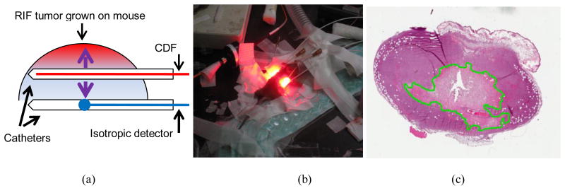
Figure 2.
The mouse necrosis experiment: (a) Schematics of treatment catheter and isotropic detectors used to measure the light fluence rate, tissue optical properties, and photosensitizer drug concentrations; (b) A picture of mice undergoing PDT; (c) The resulting tumor H&E staining slides with the necrosis edge delineated.

