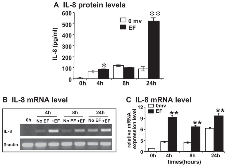
Fig. 2.
A small EF induces production of angiogenic factor IL-8 from endothelial cells. (A) IL-8 protein level; (B) semi-quantitative RT-PCR with β-actin as internal control; and (C) quantification of mRNA level. HUVEC cells were cultured in serum-free DMEM and exposed to an EF of 200 mV mm−1. IL-8 protein in the medium was quantified by ELISA. Isolated total RNA from control and EF-stimulated HUVEC were analyzed by semi-quantitative RT-PCR: histogram indicating the mRNA expression of IL-8 normalized to βactin (expressed as fold change relative to the 0 h control). IL-8 protein and mRNA transcription were increased significantly at the time points 4 and 24 h, and 4–24 h, respectively (up to two- to threefold increase compared with control). The error bars represent the SE (*p < 0.05 and **p < 0.01).

