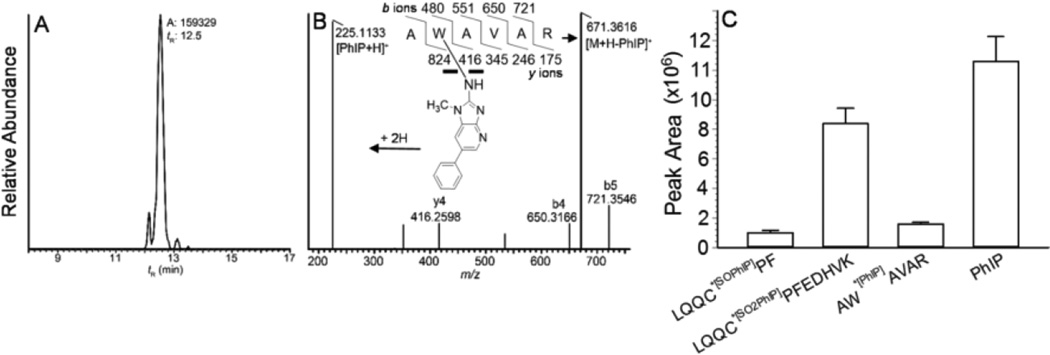
Figure 5.
(A) Reconstructed mass chromatograms obtained by CID of AW*[PhIP]AVAR ([M+2H]2+ m/z 448.2385 > 225.1135, 671.3624; mass tolerance, 2 ppm) of N-sulfooxy-PhIP modified albumin digested with trypsin, (B) product ion spectrum AW*[PhIP]AVAR, and (C) relative peak area ion counts of peptide adducts of albumin modified with N-sulfooxy-PhIP: AW*[PhIP]AVAR recovered from trypsin digest; LQQC*[SOPhIP]PF ([M+2H]2+ at m/z 487.2 > 225.1, 749.4) and PhIP ([M+H]+ at m/z 225.1 > 210.0) recovered from trypsin/chymotrypsin digest; and LQQC*[SO2PhIP]PFEDHVK ([M+3H]3+ at m/z 533.2 > 670.2, 679.0) recovered from trypsin/chymotrypsin digestion following m-CPBA oxidation of albumin.

