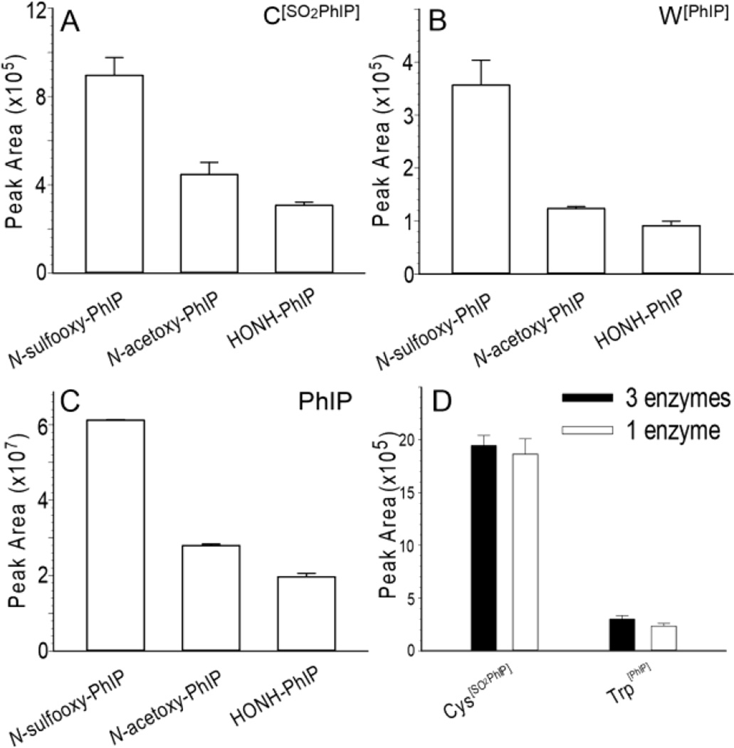
Figure 7.
(A) Peak area ion count estimates of the UPLC-ESI/MS2 analysis of (A) C[SO2PhIP] ([M+H]+ at m/z 376.1 > 225.1), (B) W[PhIP] ([M+H]+m/z, 421.1 > 225.1), and (C) PhIP ([M+H]+ at m/z 225.1 > 210.0) recovered from albumin in human plasma modified with N-sulfooxy-PhIP following by protein digestion with pronase E, leucine aminopeptidase and prolidase. (D) Peak area ion count estimates of the UPLC-ESI/MS2 analysis of C[SO2PhIP] ([M+H]+m/z, 376.1 > 225.1) and W[PhIP] ([M+H]+, m/z, 421.1 > 225.1) recovered from N-sulfooxy-PhIP modified commercial albumin following digestion with pronase E, leucine aminopeptidase and prolidase (black bar), or pronase E alone (white bar). Albumin digest (200 ng) was injected on column for all samples (Mean ± SD, n=3).

