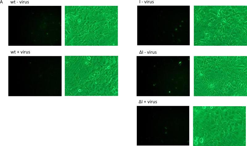
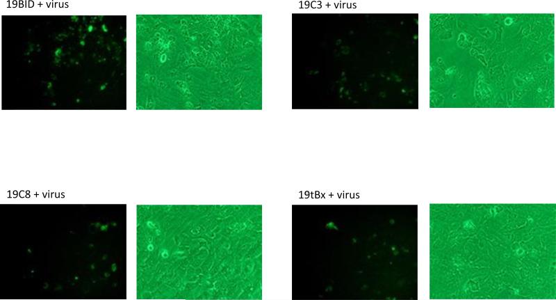
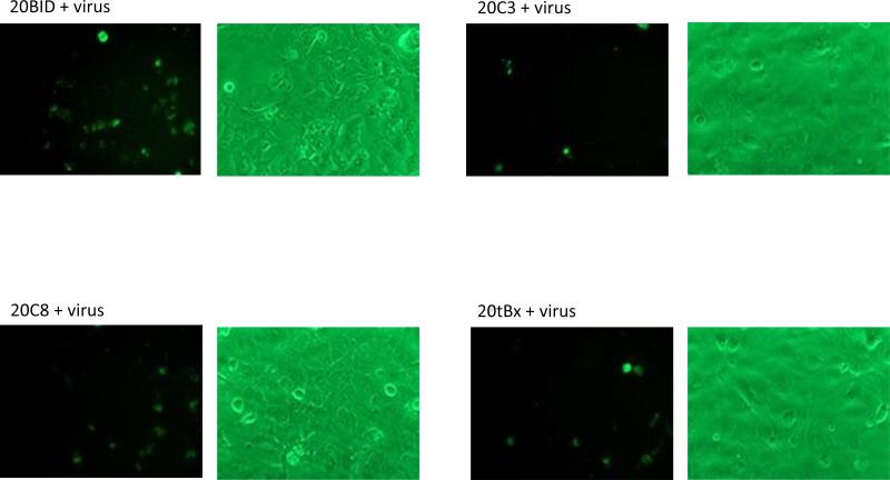
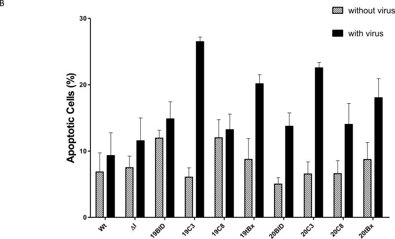
Figure 7.
Apoptotic cell death visualized by Annexin V staining. (A) HCV-infected cells were stained with Annexin V at 48 h post infection. Left: cells visualized under a fluorescent filter; Right: under a bright field; (B) Positive Annexin-V-stained cells were counted and divided by total cell count. For each sample, cells were counted manually from 4 separate fields; Wt: wild-type cells; ΔI: intron lacking a trans-splicing domain; + virus: with virus; - virus: without virus.


