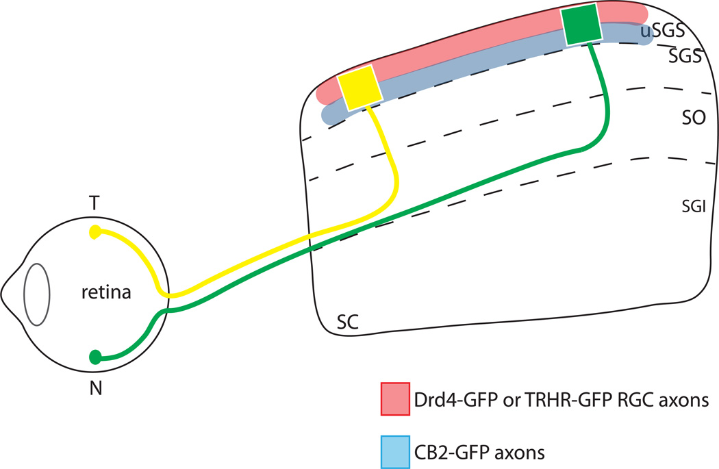
Figure 1. Anterior-posterior topography and lamination patterns of RGC types in the superior colliculus.
Temporal (T) axons of all RGC types (yellow) project to the anterior SC and nasal (N) axons (green) project to the posterior SC. Direction-selective RGC types labeled in TRHR-GFP and DRD4-GFP transgenic lines project to superficial laminae of the uSGS (upper stratum griseum superficiale) (red), Off transient RGCs labeled by CB2-GFP transgenic line project to deeper laminae of the uSGS (blue), and different types of RGCs from the same topographic location are aligned along the superficial-deep axis of the SC.

