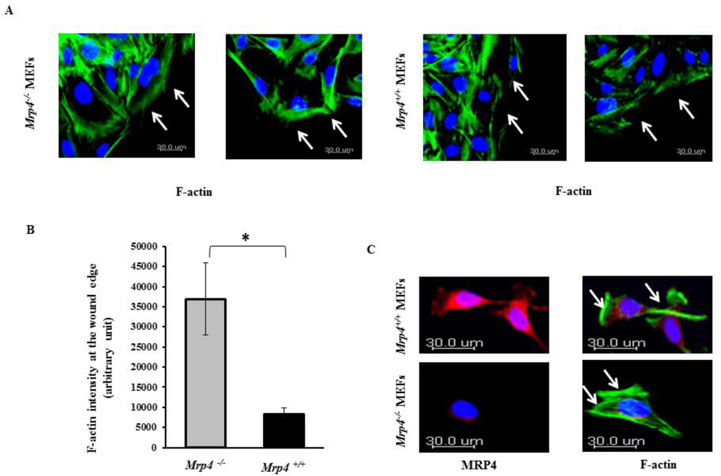
Figure 4. MRP4-deficient fibroblasts exhibit altered β-actin dynamics.
(A) Cortical actin dynamics at the wound edges by confocal fluorescence microscopy (20× objective) are shown in Mrp4+/+ and Mrp4−/− MEFs using phalloidin antibody (green). (B) Bar graph representing the total actin intensity at the wound edge in Mrp4+/+ and Mrp4−/− MEFs. Error bars indicate SEM. (C) Actin dynamics at the leading edges of migrating Mrp4+/+ and Mrp4−/− MEFs by confocal fluorescence microscopy (20× objective) are shown using phalloidin antibody (green) and anti-MRP4 antibody (red). All data represent at least three independent experiments. *, P < 0.05.

