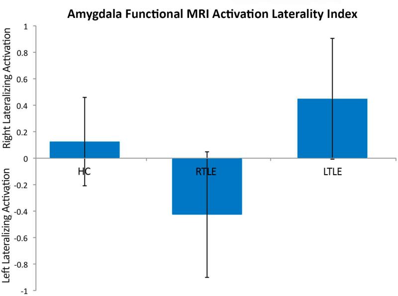
Figure 2.
Amygdala fMRI laterality index (LI). Positive LI indicating right hemisphere lateralized activation, and negative LI indicating left hemisphere lateralized activation. Individuals with TLE demonstrated a shift of amygdala activation to the side contralateral to the seizure focus in both right and left TLE.

