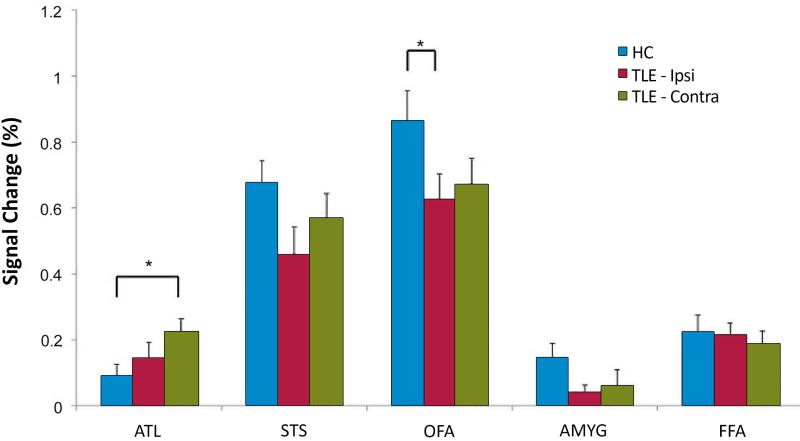
Figure 3.
Altered activation of face-responsive regions in TLE compared to healthy controls. Right and left TLE subjects were grouped together and analyses are performed according to the side of seizure onset (ipsilateral vs. contralateral). Individuals with TLE demonstrated significantly decreased activation in the ipsilateral occipital face area (OFA, *p < 0.02) and, a trend level decreased activation in the ipsilateral superior temporal sulcus (STS, p < 0.06) compared to healthy controls. Conversely, TLE subjects activated the contralateral anterior temporal lobe (ATL) significantly more than controls (*p < 0.02). Data are mean ± SEM.

