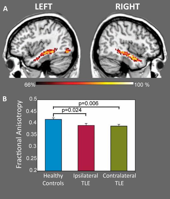
Figure 4.
White matter connectivity between face-responsive regions. Diffusion tensor imaging with probabilistic tractography delineated the inferior longitudinal fasciculus (ILF) connecting the occipital face area and anterior temporal lobe in TLE and healthy controls (top). Dark orange color indicates 66% of the group members contain these voxels and yellow color indicates 100% of the group contains these voxels within the ILF. Fractional anisotropy was significantly reduced in individuals with TLE, both ipsilateral (p = 0.024) and contralateral (p = 0.006) to side of seizure onset, compared to healthy controls (averaged across both hemispheres).

