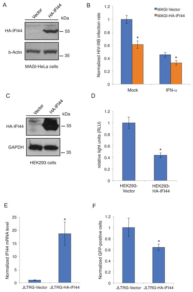
Figure 2.
Exogenous expression of IFI44 suppresses HIV-1 viral gene expression. (A). pQCXIP-HA-IFI44 or pQCXIP empty vector was transduced into MAGI-HeLa cells. Cell lysate was prepared and analyzed for HA-IFI44 expression by western blot using an anti-HA antibody. (B). MAGI-HeLa cells stably expressing HA-IFI44 or empty vector were pretreated with IFN-α or mock-treated for 24 hours. Cells were infected with HIV-IIIB wild-type viruses for 48 hours and stained with an anti-HIV-1 p24 CA antibody (anti-CA) and Hoechst. The percentage of infected cells was measured and normalized to the vector-only cells. (C). pQCXIP-HA-IFI44 or pQCXIP empty vector was transduced into HEK293 cells. Cell lysate was prepared and analyzed for HA-IFI44 expression by western blot using an anti-HA antibody. (D). HEK293 cells stably expressing HA-IFI44 or empty vector were co-transfected with HIV-LTR firefly luciferase reporter and pRL-TK renilla luciferase control vectors, together with pcDNA-FLAG-TAT vector. Luciferase activity was measured 24 hours post-transfection and normalized to the empty vector cells. (E). pQCXIP-HA-IFI44 or pQCXIP empty vector was transduced into JLTRG cells. Total RNA was extracted for reverse transcription and quantitative real-time PCR to measure the IFI44 mRNA level. Data were normalized to the JLTRG-Vector cells. (F). JLTRG cells stably expressing HA-IFI44 or empty vector were transiently transfected with pcDNA-FLAG-TAT. 48 hours post-transfection, cells were assessed by flow cytometry, and the percentage of GFP-positive cells was normalized to the vector-only cells. All results were presented as mean ± s.d. (n = 3); * P < 0.05 from t-test.

