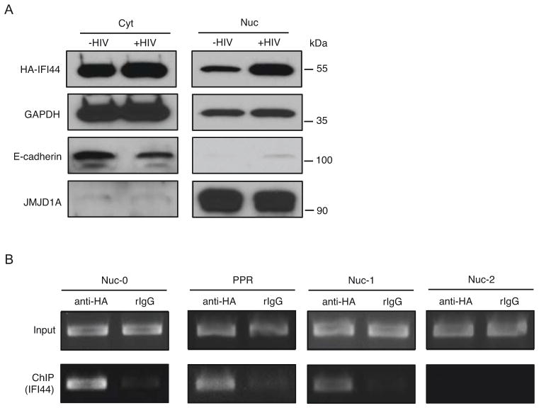
Figure 3.
IFI44 enters the nuclei and associates with the HIV-1 LTR upon HIV-1 infection. (A). HEK293 cells stably expressing HA-IFI44 were infected with VSV-G pseudo-typed HIV-NL4-3-GFP [Δ Env] viruses (+HIV, MOI=5) or mock treated (−HIV) for 48 hours. Equal numbers of cells were subjected to cytoplasmic (cyt) and nuclear (nuc) fractionation. Extracted protein samples were separated by SDS-PAGE. HA-IFI44 cyt or nuc localization was analyzed by western blot using an anti-HA antibody. GAPDH protein level in cyt or nuc extraction was determined to indicate equal loading. E-cadherin (cytoplasm) and JMJD1A (nuclei) were stained to ensure the separation of cytoplasmic and nuclear proteins. (B). HEK293 cells stably expressing HA-IFI44 were infected with VSV-G pseudo-typed HIV-1 NL4-3-GFP [Δ Env] viruses. Cells were subjected to ChIP assays using an anti-HA antibody or a rabbit control IgG. Precipitated DNA samples were extracted and analyzed by PCR using primers amplifying the nuc-0, L1 nucleosome-free region (PPR), nuc-1, and nuc-2 regions of the HIV-1 LTR promoter.

