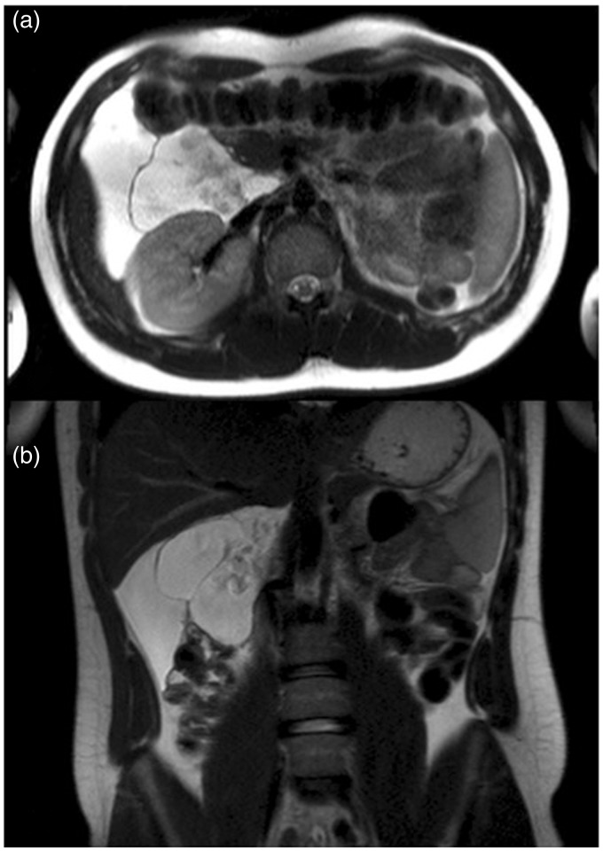
Fig. 2.
MRI of the abdomen. Axial (a) and coronal (b) T2W TSE images showed the presence of a huge fluid-filled mass with internal septa located in the hepato-renal space extending from the hepatic hilum to the right colon and displacing medially the second duodenal segment and anteriorly the hepatic flexure of the colon.

