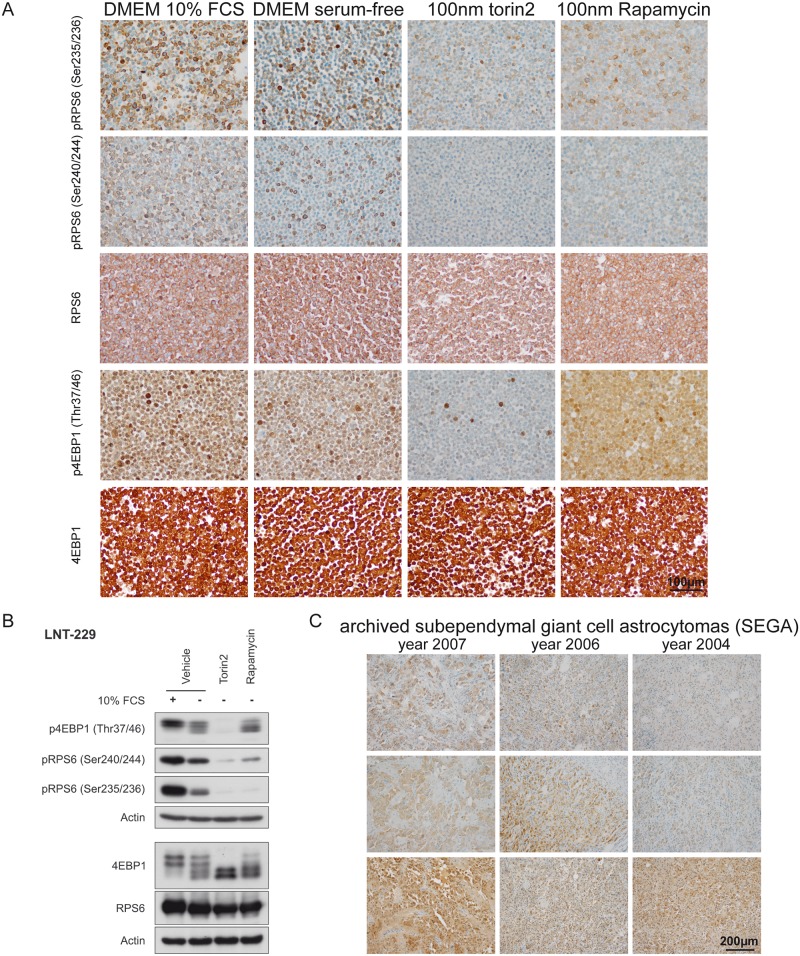
Fig 2. Establishment of mTORC1-specific immunohisto- and immunocytochemistry.
(A) LNT-229 glioma cells cultured in DMEM containing 10% FCS and 25 mM glucose displayed strong signals in phospho-specific ICC stainings with all tested antibodies, while serum-free culture condititons resulted in reduced signal intensity and frequency. Treatment with the ATP-competitive mTORC-inhibitor torin2 resulted in a profound reduction of the phospho-spcific signal for all antibodies tested, while treatment with rapamycin only reduced the signal of RPS6 phospho-sites. Total RPS6 and 4EBP1 protein counterparts did not show considerable differences in the tested conditions (B) Immunoblotting using the same antibodies, cell lines and culture conditions confirmed the ICC results presented in (A), 4EBP1 exists in states of different electrophoretic mobility depending on its phosphorylation state [19]. (C) Subependymal giant cell astrocytomas (SEGA) served as a positive control for human routine diagnostic specimens. Although mTORC1-signaling was detectable in SEGA tumor cells, the staining intensity was considerably reduced in freshly cut archived FFPE material especially for the RPS6 phospho-sites.

