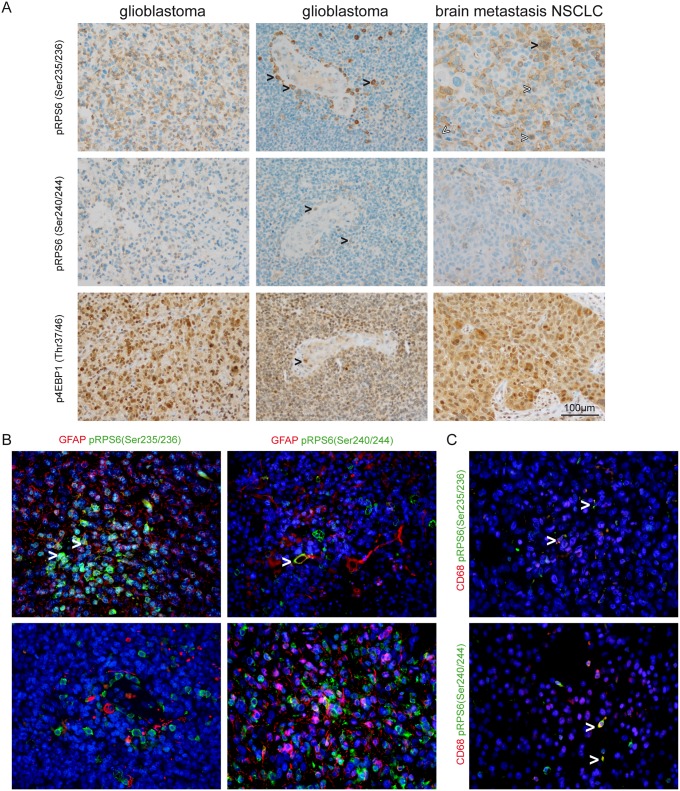
Fig 5. Detection of phospho-RPS6 (Ser235/236), phospho-RPS6 (Ser240/244) and phospho-4EBP1 (Thr37/46) in malignant brain tumors.
(A) Although pleomorphic tumor cells showed staining for phospho-RPS6 (Ser235/236) and phospho-RPS6 (Ser240/244) we also observed perivascular cells with microglia/macrophage morphology that were strongly positive for both markers (black arrowheads in the first and second row of glioblastoma). We also detected strong expression of both phosphorylated antigens in brain metastases of NSCLC tumor cells (black arrowheads in the upper row, right column indicating multinucleated tumor cells, white arrowheads indicating mitotic figures). In contrast to the more heterogenous staining for phospho-RPS6 (Ser235/236) and phospho-RPS6 (Ser240/244), phospho-4EBP1 (Thr37/46) was strongly detected in the majority of tumor cells. Vessel-associated cells are also a source for mTORC1-signaling (black arrowhead indicating a mitotic figure within the endothelial layer, lower middle image). (B) Immunofluorescent double staining of glioblastomas against phospho-RPS6 (Ser235/236) and (Ser240/244) showing colocalisation with GFAP in some tumor cells (white arrowheads) while numerous GFAP-positive cells did not show phospho-signals for RPS6. (C) Phospho-RPS6 (Ser235/236) and (Ser240/244) were detected in CD68-positive cells in glioblastomas (white arrowheads).

