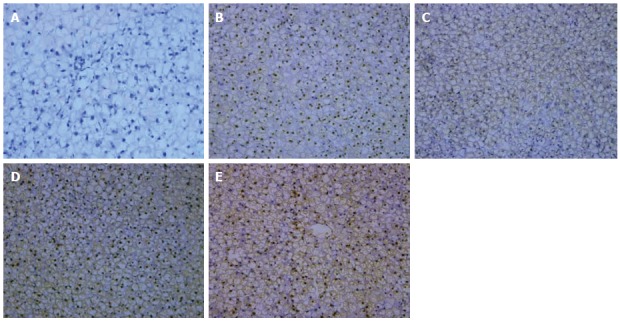
Figure 2.

Immunohistochemical staining for nuclear factor-κB protein expression (SP, magnification × 200). A: The control group; B: The model group at the end of the 6th week; C: The model group at the end of the 10th week; D: The model group at the end of the 14th week; E: The intervention group at the end of the 14th week.
