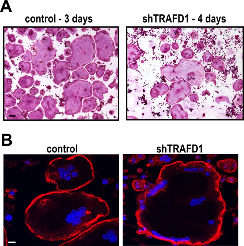
Fig 5. shTRAFD1 cells differentiate in vitro.

(A) Cells stably expressing TRAFD1 shRNAs were cultured on 96-well plates in the presence of RANKL (10 ng/ml), fixed, and stained for TRAP on the days indicated. Representative micrographs are shown. Scale bar = 100 μm. (B) Rhodamine phalloidin and DAPI staining of osteoclast-like cells cultured on glass coverslips. shTRAFD1 and control RAW264.7 cells were cultured in the presence of RANKL (10 ng/ml) on 24-well plates with glass coverslips on the bottom. When cells differentiated (3 days for controls, 4 days for shTRAFD1), they were fixed and stained. Representative fluorescence micrographs are shown. Scale bar = 10 μm.
