Fig. 1.
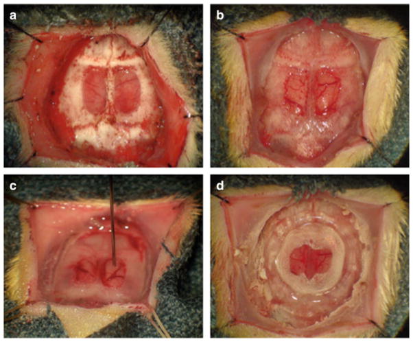
Essential stages in the creation of a brain tumor window model: After reflecting the scalp edges laterally, a nearly full thickness craniectomy is performed, sparing the bone over the superior sagittal sinus (a). Once the remaining bone overlying the dura has been removed, a small durotomy at the base of the craniectomy is created to create a larger linear dural rent which is used to peel the dura forward, ideally in a single flap. Once the dural flap has been removed, the shiny arachnoidal surface of the brain is appreciated (b). Under microscopic guidance, a 10 μL, 26 gauge Hamilton syringe mounted to a steretactic injector is lowered through the meninges, 1 mm deep into an avasacular region of cortex (c). After injection, the surface of the brain is irrigated to remove residual tumor cells. A 10 mm glass cover slip is carefully placed on the surface of the brain and a confluent circumferential layer of cyanoacrylate glue, overlapping the edge of the coverslip and the craniectomy margin is applied (d). Glue is usually dry within 45 min at which time a plastic ring stenting the scalp open for continuous observation is applied

