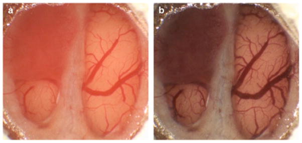
Fig. 3.

Images of a brain tumor window model 12-days post-implantation before (a) and 50 min after Coomassie blue administration (b). Note the relatively subtle difference in the color of the tumor prior to contrast administration and the clear difference between the appearance of tumor and normal brain post-contrast
