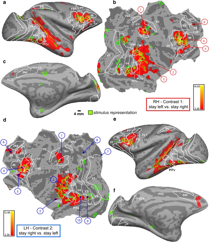
Figure 3.
Contralateral modulation of attention. FreeSurfer F99 inflated (a,e, lateral view; c,f, medial view) and flattened (b,d) surfaces, displaying MFX contrasts 1 and 2 (p = 0.001, t = 3.39). a–c, Right hemisphere (RH): stay left versus stay right. d–f, Left hemisphere (LH): stay right versus stay left. Green transparent overlay, Activation of the stimulus localizer experiment (MFX contrast: bilateral stimulus vs fixation thresholded at p = 0.01, t = 2.45). Sulci: sts, Superior temporal; ips, intraparietal; cs, central; pos, parieto-occipital; cing, cingulate. Numbers indicate areas for which example time courses are plotted in Figures 4, 5, 6, and 9. White outlines indicate areal boundaries based on retinotopy labels (probability maps of 5 monkeys, including M13, M24) (Janssens et al., 2014): V1–V4 (A), posterior inferotemporal dorsal (PITd), PITv (ventral), and middle temporal (MT) (Nelissen et al., 2011): F5, F6, and F7, as well as LIP, FEFs, V6A (Lewis and Van Essen, 2000b): 24 d, 45, 46, 5V (PE).

