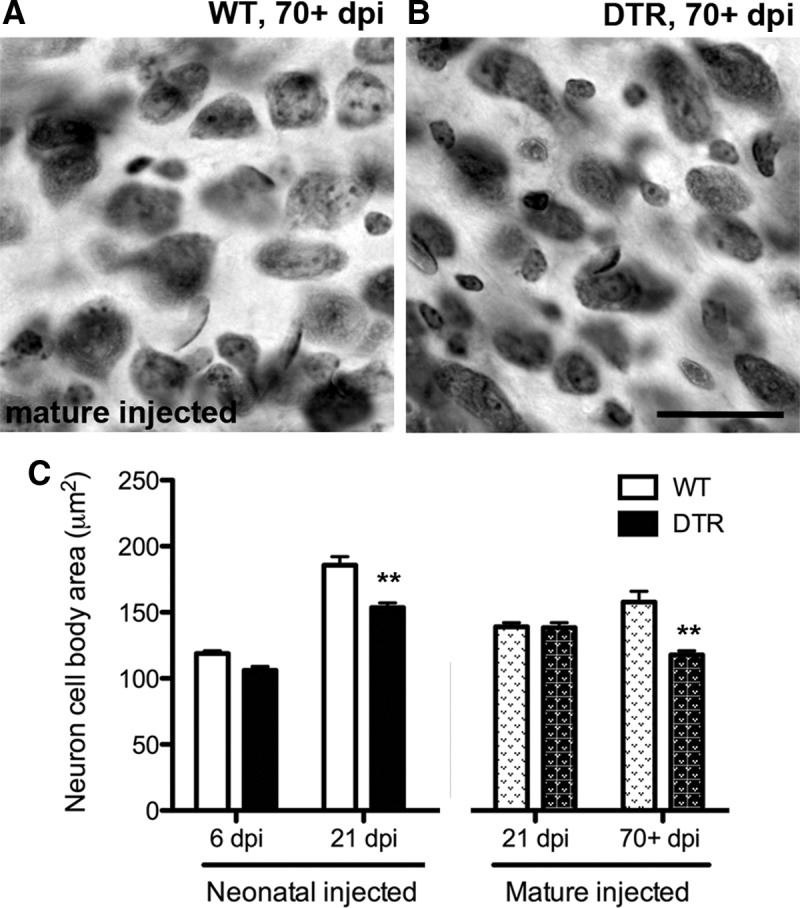
Figure 12.

Neuronal cross-sectional area decreases slowly after cochlear hair cell loss. A, B, Photomicrographs of section through the middle region of the ventral CN of mature WT and DTR mouse, respectively, 70+ dpi. Note that a small difference in neuron size is apparent, with soma area slightly smaller in DTR mouse CN. Scale bar, 25 μm. C, Cross-sectional area measurements of WT and DTR mice at various survival times. In neonatal mice injected with DT, neuronal size in the surviving neurons is significantly decreased by 21 dpi. A small, but significant, decrease is also detected when mature mice are injected with DT, but on a more delayed time course; Data are mean ± SEM, n = 3–4; **p < 0.001 (vs age-matched controls); two-way ANOVAs followed by Bonferroni post hoc tests.
