
Figure 6.
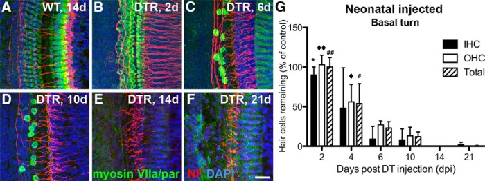
Cochlear hair cells are lost quickly after injection of DT in neonatal (P2) DTR mice. All data are from WT and DTR mice injected with DT at P2 with 4 ng/g body wt. A, Hair cells (green) remain largely intact in WT mice after injection of DT, even after long survival periods (shown here 14 dpi). B, Hair cells are swollen and unhealthy looking in DTR mice as early as 2 dpi. C, D, At 6 and 10 dpi, extensive loss of both inner and outer hair cells is apparent, with almost complete loss of inner hair cells and the remaining outer hair cells appearing pathologically swollen. E, F, At 14 and 21 dpi, all hair cells are gone in the basal turn, but a few hair cells (<5%) remain in more apical regions (Table 2). Nerve fibers from SGNs (red) are also markedly reduced after substantial hair cell loss, and this was especially apparent by 14 dpi and beyond. Scale bar, 25 μm. G, Graph displays loss of inner, outer, and total hair cells in the middle of the basal turn of DTR mice. Data are mean ± SEM (n = 3 cochleas from different mice). Statistical analyses (one-way ANOVA followed by Bonferroni post hoc tests) yielded significant effects of time dpi: *p < 0.05 (2 vs 6, 10, 14, and 21 dpi); ♦♦ p < 0.05 (2 dpi vs all others); ## p < 0.05 (2 dpi vs all others); ♦ p < 0.05 (4 vs 6, 10, 14, and 21 dpi); # p < 0.05 (4 vs 6, 10, 14, and 21 dpi).