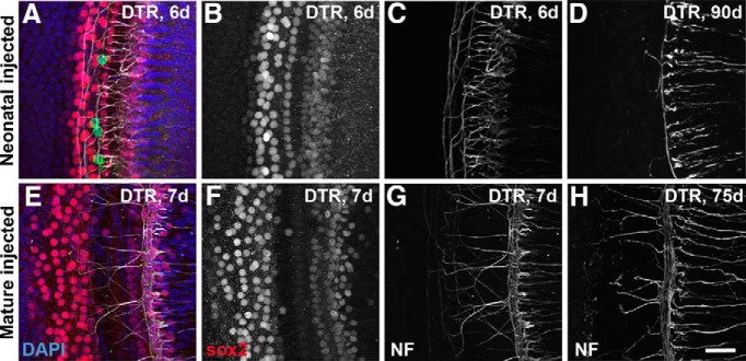
Figure 7.
Effect of DT on supporting cells and peripheral neurites in the cochlea of DTR mice. A–C, Confocal images from a single confocal stack taken from a DTR mouse injected with DT on P2 and allowed to survive 6 d (middle turn shown here). A, Merged image with Sox2 labeling of organ of Corti supporting cell nuclei (red), neurofilament reactivity (white), and DAPI nuclear counterstain (blue) and hair cells labeled with myosin VIIa (green). Note that, whereas hair cells are virtually completely gone, supporting cells appear unaffected at short survival times. B, Sox2 channel alone. C, Neurofilament channel alone. D, Neurofilament labeling in the same region from a DTR mouse injected with DT at P2, but allowed to survive 90 d; note extensive loss of neurites and virtual absence of neurites crossing to the outer hair cell region. E–G, Confocal images from a single confocal stack taken from a mature DTR mouse injected with DT on P40 and allowed to survive 7 d (apical turn shown here). E, Merged image labeled as in A. F, Sox2 channel alone. G, Neurofilament channel alone. H, Neurofilament labeling in the same region from a DTR mouse injected with DT at P40, but allowed to survive 75 d; note extensive labeling of neurites that is essentially as dense as seen after only 7 d survival (G) and as seen in WT mice. Also note normal numbers of neurites crossing to the outer hair cell region. These tissues were also labeled with an antibody against myosin VII for hair cells. Four hair cells (green) are seen in A. All other hair cells have been eliminated by the DT. Scale bar, 25 μm.

