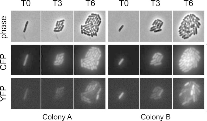
FIG 5.
Time-lapse microscopy of two representative microcolonies. Cultures of strain MMR8 [PtorCAD-yfp Φ(ompA+-cfp+)] were grown aerobically in minimal medium with 10 mM TMAO. Cells were immobilized on 5-mm-thick agarose pads composed of 1% agarose dissolved in the same growth medium with TMAO and maintained on the microscope stage at 37°C with exposure to air. The images shown are of two representative colonies, each derived from a single cell, shortly after immobilization on agarose pads (T0) and then 3 h (T3) and 6 h (T6) after the starting time point (approximately four and eight divisions, respectively). The rows consist of phase-contrast images (top), CFP fluorescence images (middle), and YFP fluorescence images (bottom).

