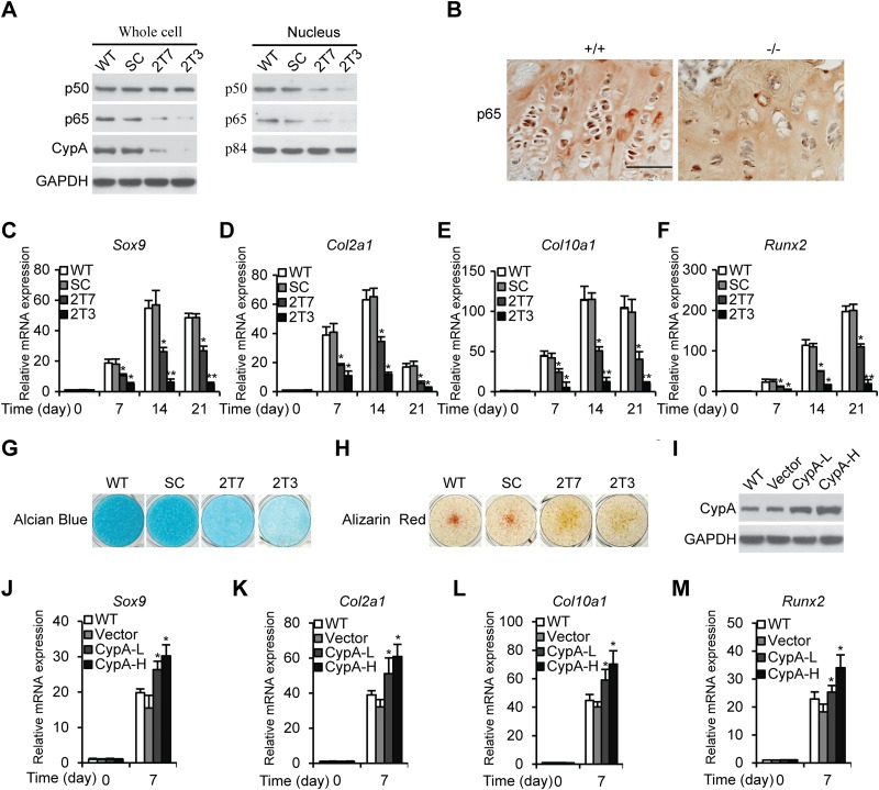
FIG 1.
Knockdown of CypA attenuates chondrogenic differentiation of ATDC5 cells. (A) Western blot analyses. Whole-cell lysates and nuclear extracts prepared from ATDC5 WT, SC, and CypA Kd stable clones 2T7and 2T3 were immunoblotted to detect CypA, p65, and p50 expression levels. GAPDH and p84 were used as loading controls. (B) Immunohistochemical staining of p65 in a dewaxed section prepared from the distal femoral growth plates of 3-week-old WT and CypA KO mice (scale bar, 50 μm). (C to F) Chondrogenic differentiation of ATDC5 WT, SC, and CypA Kd clones 2T7 and 2T3. The relative mRNA levels of Sox9 (C), Col2α1 (D), Col10α1 (E), and Runx2 (F) were measured by RT-quantitative PCR at several time points. Mean values (n = 3) and standard deviations (SD) are shown. *, P < 0.05, and **, P < 0.01 compared to WT cells at the same time points. (G and H) Alcian blue (G) and alizarin red (H) staining of cell surface proteoglycans and calcium deposits from 21-day chondrogenic differentiated ATDC5 WT, SC, and CypAKd clones. (I) Western blot analyses of CypA expression levels. WT, wild-type ATDC5 cells; Vector, empty-vector-transfected WT cells as a control; CypA-L, WT cells transfected with low concentrations of CypA plasmids (1.25 μg/well) in 6-well plates; CypA-H, WT cells transfected with high concentrations of CypA plasmids (2.5 μg/well) in 6-well plates. (J to M) Relative mRNA expression levels of Sox9 (J), Col2α1 (K), Col10α1 (L), and Runx2 (M) in ATDC5 WT, vector-transfected, and CypA-L-transfected or CypA-H-transfected ATDC5 cells cultured in chondrogenic differentiation medium for 21 days. The data are shown as means and SD of three experiments. *, P < 0.05 compared to WT cells at the same time points.

