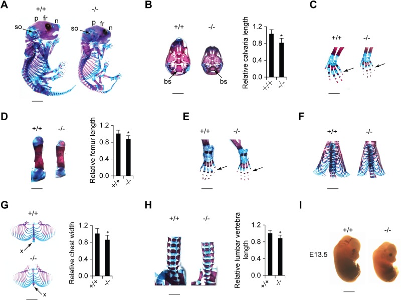
FIG 5.
Ppia−/− mice exhibit small body size and defects in skeletal development. (A) Alcian blue/alizarin red staining of skeletons of newborn Ppia+/+ and Ppia−/− mice (scale bar, 3 mm). A lateral view of the skull exhibits differences in the development of the frontal (fr), parietal (p), nasal (n), and supraoccipital (so) bones. (B) (Left) Ventral view of the caudal part of the skull showing that the basisphenoid bone (bs) is smaller in the Ppia−/− newborn mouse (scale bar, 2 mm). (Right) Quantification of skull length. (C) Left forelimbs of newborn mice (scale bar, 1 mm). The arrows indicate the distal phalanges of the fifth digit of the forepaws. (D) Comparison of left femurs of Ppia+/+ and Ppia−/− mice with double staining (left) (scale bar, 2 mm) and length quantification (right). (E) Double staining of left hind limbs (scale bar, 1 mm). The arrows indicate the distal phalanges of the fifth digit of the hind paws. (F) Ventral view of backbones and rib cages (scale bar, 3 mm). (G) Rib cages (left) (scale bar, 3 mm) and width quantification (right). The arrows indicate xiphoid processes. (H) Frontal view of the lumbar vertebrae with alcian blue and alizarin red staining (left) (scale bar, 2 mm) and length quantification (right). (I) Comparison of the sizes of Ppia+/+ and Ppia−/− embryos at E13.5 (scale bar, 1 mm).

