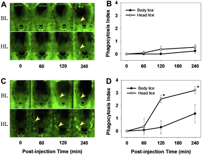
Fig. 4.
Fluorescence microscopic images of abdominal region of the lice injected with FITC-labeled Staphylococcus aureus (A) or Escherichia coli (C) and the time course of phagocytosis as determined by phagocytosis index (B and D). Yellow arrows in the panels A and C indicates the typical phagocytes clusters immobilized in the lateral region of abdomen. The size of body louse images was reduced 1.5 fold to make it similar to that of head lice. BL, body lice; HL, head lice.

