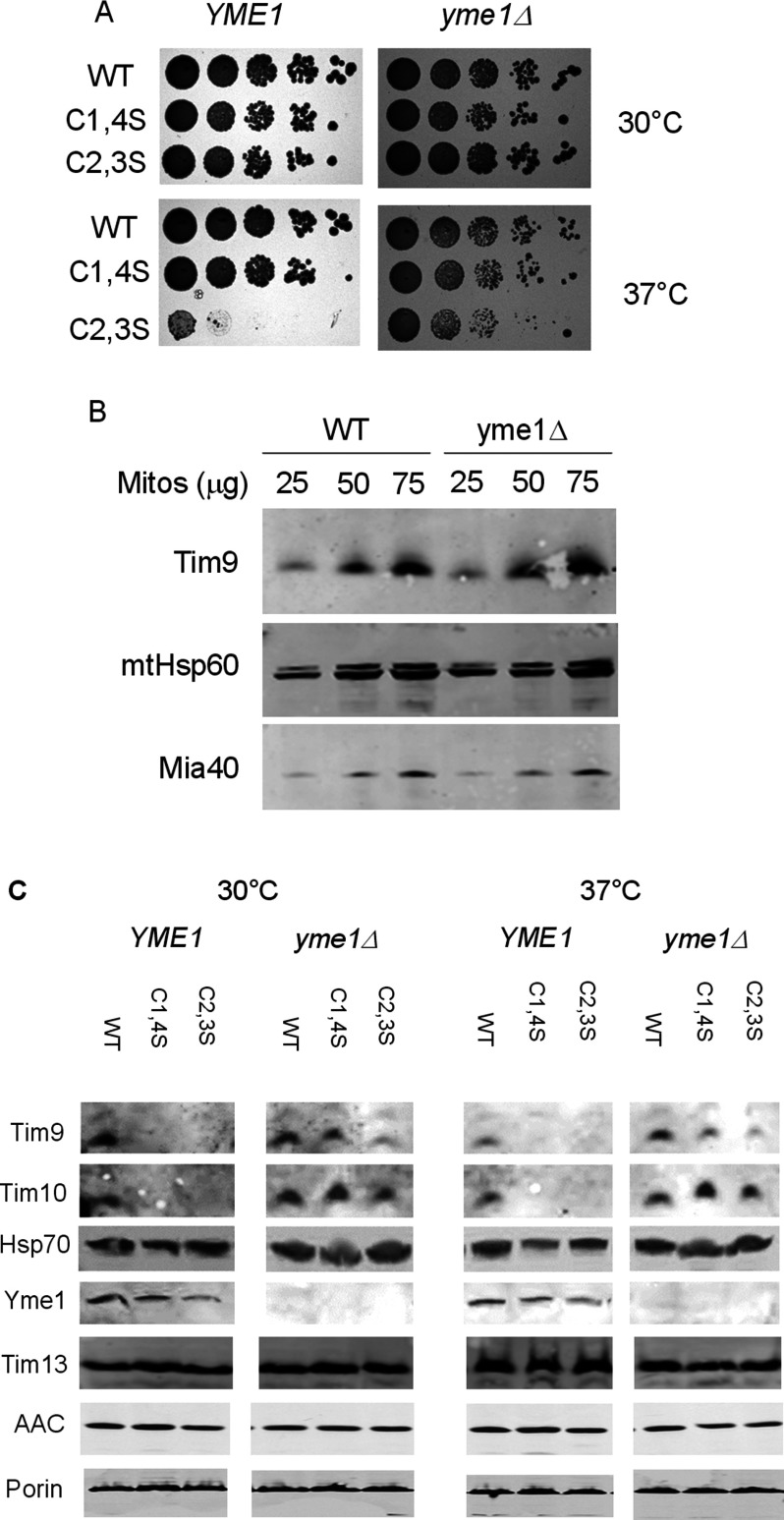
Figure 2. Deletion of YME1 rescues tim9C2,S growth defect and Tim10 mitochondrial levels.
(A) Spot tests for growth of tim9 mutants in the yme1Δ background on YPD media at 30°C and 37°C. (B). Western blot for levels of mitochondrial proteins in YME1 and yme1Δ mitochondria. Mitochondria were lysed and proteins detected with the indicated antibodies (C). Western blots for levels of mitochondrial proteins in WT and tim9 mitochondria in the presence or absence of Yme1. Isolated mitochondria were pre-incubated at either 30°C or 37°C for 15 min and then lysed. Fifty microgram was separated by SDS/PAGE and protein levels were detected using the indicated antibodies.

