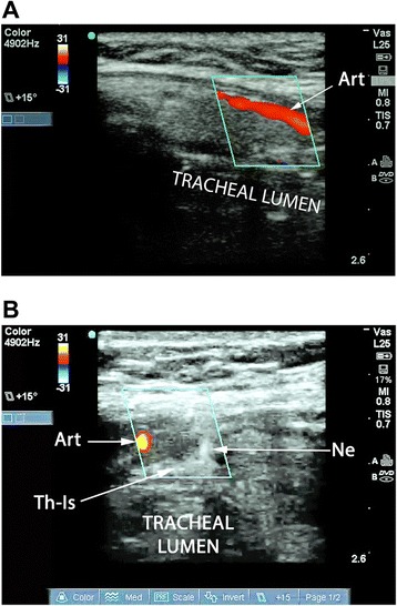
Figure 5.

Visualization and avoidance of vascular structures. (A) Longitudinal view of the trachea with duplex imaging. A paramedian artery is seen, likely the thyroid ima. (B) Axial view of the trachea during puncture with duplex imaging (same patient as in (A)). The needle tip is directed to the anterior tracheal wall under real-time duplex guidance while avoiding the previously seen paramedian artery (likely the thyroid ima). Art, artery; Ne, needle tip; Th-Is, thyroid isthmus.
