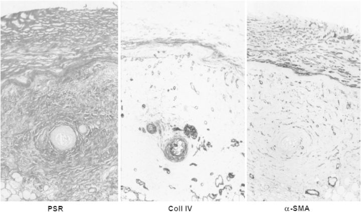
Fig. 6.
The same section of parietal peritoneum of a patient with encapsulating peritoneal sclerosis, stained with picosirius red (stains fibrillary collagen), anticollagen IV (stains basement membranes) and α-smooth muscle actin (stains myofibroblasts). From: Mateijsen MAM et al. Perit Dial Int 1999;19: 517–525.

