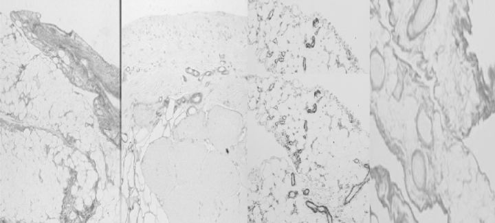
Fig. 7.
The morphology of the peritoneum of rats exposed to a conventional dialysis solution for 20 weeks. Left panel: parietal peritoneum, picosirius red. Mid-left: parietal peritoneum, antifactor VIII staining. Mid-right: omentum: α-smooth muscle actin. Right panel: omentum, picosirius red. Note the similarities with the studies in humans (Figs 5 and 6).

