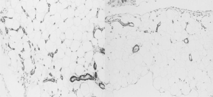
Fig. 9.
Omental tissue of a rat stained with α-smooth muscle actin after 20 weeks exposure to a conventional lactate-buffered dialysis solution (left panel) and after 20 weeks exposure to a similar, but pyruvate-buffered dialysis solution (right panel). Note the differences in the number of blood vessels and the amount of fibrosis.

