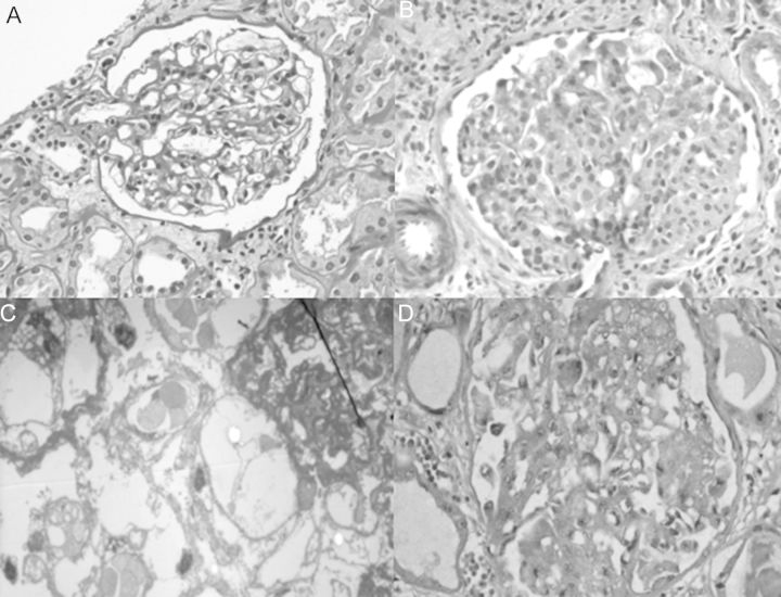
Fig. 1.
Photomicrograph showing (A) normal glomeruli by LM (PAS, ×40), and complete foot process effacement by EM (not shown), consistent with minimal-change disease; (B) Segmental collapse with overlying proliferation of visceral epithelial cells consistent with collapsing glomerulopathy (Masson's trichrome, ×40), (C) electron microscrograph shows hypertrophied podocytes with vacuoles over a collapsed glomerular tuft (uranyl acetate, ×62 000) and (D) proliferating visceral epithelial cells stained with Ki67 (IHC, ×40)

