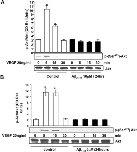
Figure 6.

Representative Western blotting and densitometric analysis showing Akt phosphorylation at serine 473 in BAECs after stimulation with VEGF 20 ng/ml for 0 to 30 min. BAECs were cultured for 24 h in the presence (black bars) or absence (control, white bars) of 10 μM Aβ25–35 (A) and 5 μM Aβ1–42 (B). Data were analyzed for statistical significance (*P < 0.01 versus 0 min control group, n = 3). VEGF, vascular endothelial growth factor.
