
Figure 2.
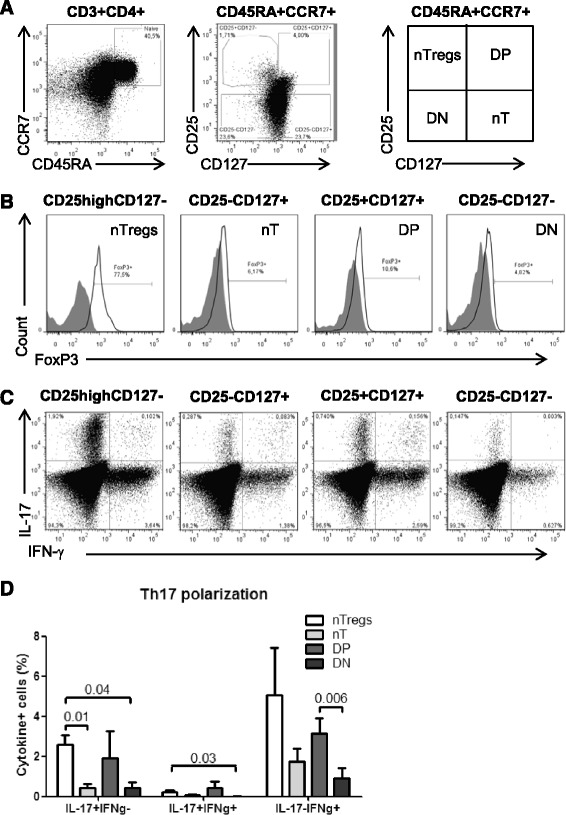
Phenotypically naive CD25highCD127−FoxP3+ and CD25+CD127+FoxP3− T-cells preferentially differentiate into Th17 cells. PBMCs from HIV- controls were stained with a cocktail of CD3, CD4, CD45RA, CCR7, CD25 and CD127 Abs on the surface and intracellularly with FoxP3 Abs. (A) The differential expression of CD25 and CD127 distinguished four CD45RA+CCR7+ CD4+ T-cells subsets: CD25highCD127− (nTregs), CD25−CD127+ (nT, conventional), CD25+CD127+ (DP, double positive) and CD25−CD127− (DN, double negative). (B) Shown is the expression of FoxP3 on these four subsets. (C-D) The four CD45RA+CCR7+ T-cell subsets were sorted by flow cytometry as in Additional file 2: Figure S2, stimulated via CD3/CD28, and cultivated under Th17 polarizing conditions and assessed for the co-expression of IL-17A and IFN-γ as in Figure 1. Vivid staining was used to exclude dead cells. (A-C) Results are from one donor representative of experiments performed with cells from n = 4 donors. (D) Shown are statistical analysis of Th17-polarized nTregs (n = 4), nT (n = 4), DP (n = 3), and DN cells (n = 3) with differential expression of IL-17A and IFN-γ. Paired t-Test p-values are indicated on the graph. Clinical parameters of subjects included in these studies are included in Table 1 (HIV- # 3, 5, 6, 15).