
Figure 1.
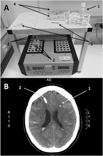
Materials and methods. A: Setup for in vitro microdialysis. 1, cerebrospinal fluid sample; 2, identical aliquots of the cerebrospinal fluid; 3, two microdialysis catheters with a membrane length of 10 mm and a 20 kDa cut-off; 4, perfusion pumps; 5, vials gathering samples of microdialysis fluid. B: Computed tomography scan from SAH patient 3. 1, a microdialysis catheter placed in the left frontal lobe; 2, part of the external drain passing through the brain.