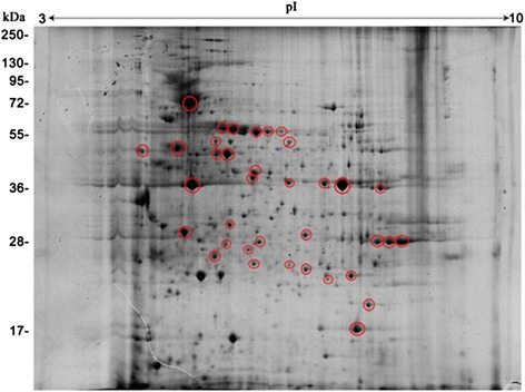
Fig. 3.

Antigenic proteins on corresponding representative 2DE gels of the proteins expressed by E. granulosus adult worms. Proteins (800 μg) were separated on a linear pH range of 3–10 by using IEF in the first dimension and 12 % SDS-PAGE in the second dimension. Proteins were electrotransferred to PVDF membranes and probed with sera from E. granulosus-infected dogs. Antigenic protein spots are indicated by red circles. Molecular weight markers are shown on the left. The proteins identified are shown in detail in Table 2
