
Fig. 3.
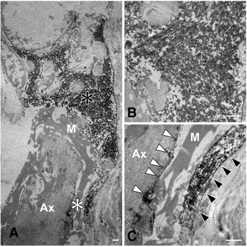
Immunoelectron microscopy of Schwann cell cytoplasmic inclusions in the spinal nerve roots. a Phosphorylated α-synuclein-immunoreactive structures in the cytoplasm of Schwann cells. b A higher-magnification view of the area indicated by the black asterisk in (a). The inclusion showing granule-coated fibrillary structures, about 15–20 nm in diameter. Anti-phosphorylated α-synuclein antibody labels filamentous and granular structures. c A higher-magnification view of the area indicated by the white asterisk in (a). Phosphorylated α-synuclein-immunoreactive structures are evident in the outer (black arrowheads) and inner loops (white arrowheads) of Schwann cells. M, myelin; Ax, axon. Bars = 1 μm