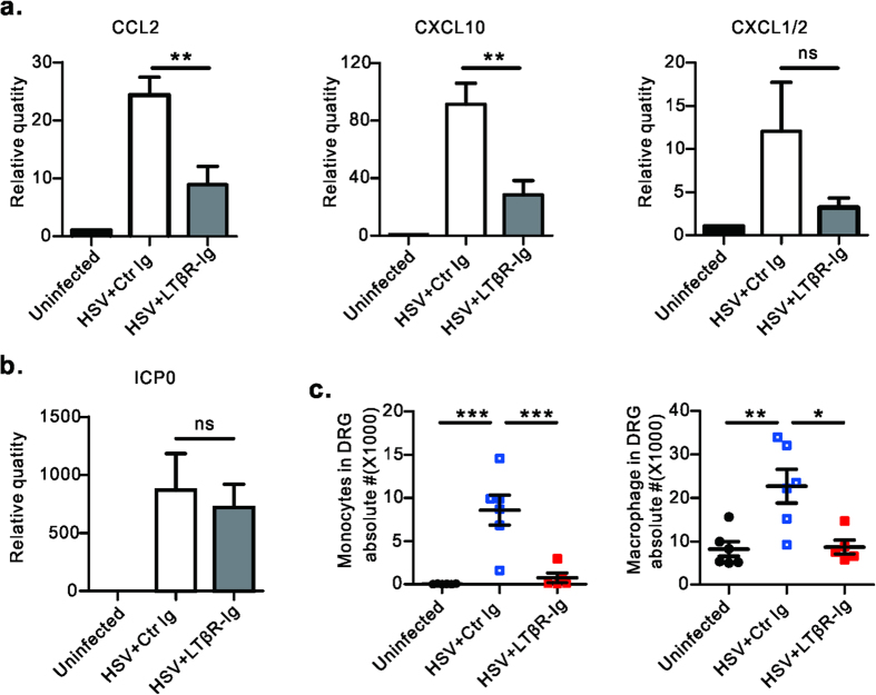
Figure 4. Blockade of LT/LIGHT inhibits chemokine expression and inflammatory cell infiltration into the DRG of infected Rag1–/– mice.
Rag1–/– mice (n = 3 to 5/group) were infected with HSV-1 and treated with LTβR-Ig as Fig. 1a. Uninfected mice were chosen as the control group. On day 6 p.i., DRGs (L3, L4 and L5) were collected from euthanized mice. The mRNA level of various chemokines (CCL2, CXCL10 and CXCL1/2) (a) and viral ICP0 gene (b) were measured by real-time PCR. Data are representative of two independent experiments. (c) On day 8 p.i., innate immune cell subsets in DRG were determined by flow cytometry assay. Gate strategy: monocytes (CD45+CD11b+Ly6ChiLy6Gmiddle), macrophage (CD45+F4/80+). Data are pooled from two independent experiments, n = 5 to 6/group. Statistical analysis for a, b, c was by unpaired t test. Error bar represents SEM, *p < 0.05, **p < 0.01, ***p < 0.001; ns, no significant difference.

