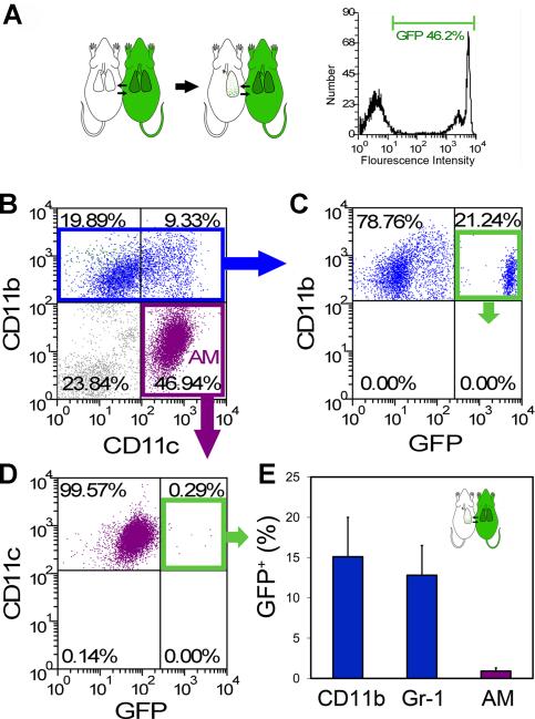
Figure 4.
Parabiotic demonstration of rare blood-borne AM after pneumonectomy. A) Schematic of the parabiotic experiments with parabiosis being established for 28 days prior to left pneumonectomy (left). Cross-circulation equilibrium was confirmed by single parameter flow cytometry (right). B-D) Because the cumulative effects of cell activation, migration and proliferation were maximal on day 14, BAL cells were studied 14 days after pneumonectomy. B) After gating CD11b+ Gr-1- (blue) or CD11b- CD11c+ cell populations (purple), the frequency of GFP+ cell migration was determined. Representative dual parameter histograms are shown (C, D). E) Flow cytometry of BAL cells demonstrated that an average of 15.1+4.9% of CD11b+ Gr-1- cells, 12.8+3.7% of CD11b+ Gr-1+ cells and 0.9+0.4% of AM were blood-borne cells (p<.001; N=4 mice, error bars reflect the mean + 1 SD). Of note, separately analyzed AM from the GFP+ parabiont demonstrated GFP expression similar to the CD11b+ cells shown here (C).

