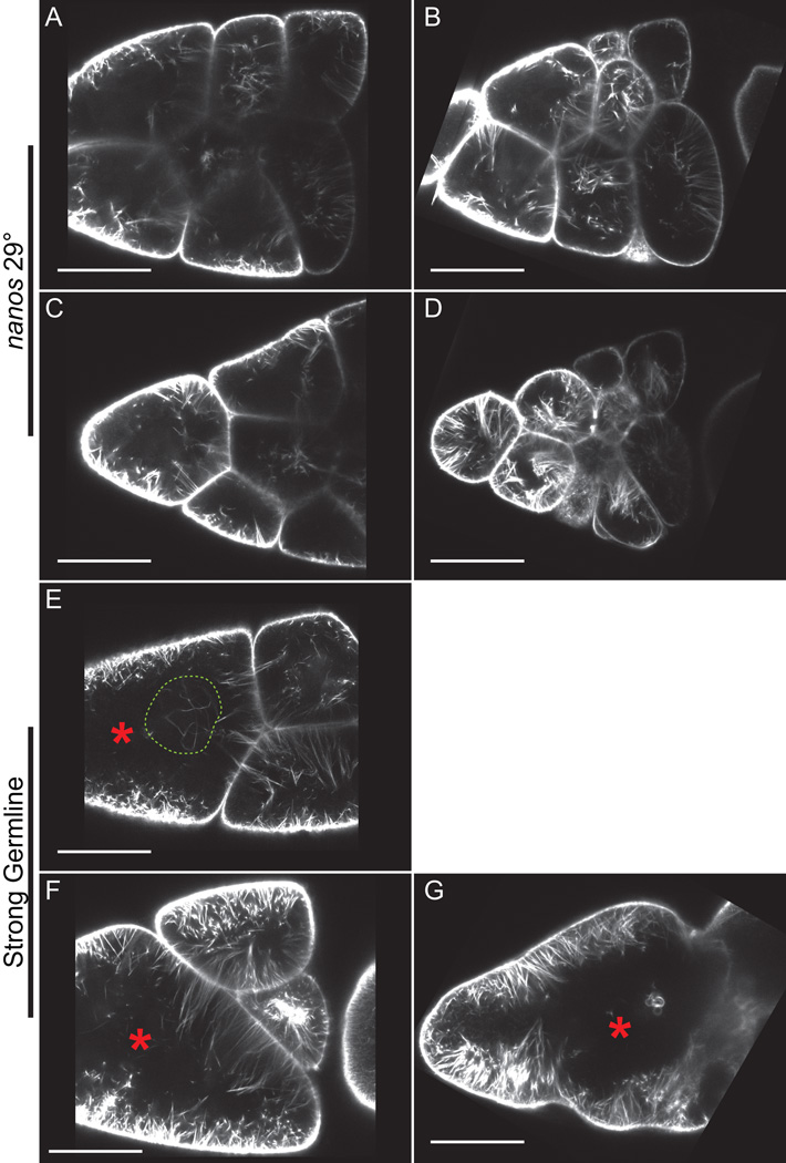
Figure 4. Live imaging of S10B GFP-Utrophin expressing follicles.
(A-G) Single focal plane from confocal z-stack of live S10B-S11 follicles taken at 40X magnification. (A-G) GFP-Utrophin = white. (A-D) GFP-Utrophin; nanos-VP16 GAL4 at 29°C. (E-G) Strong germline GAL4 (mat3 or oskar GAL4) driving GFP-Utrophin (room temperature). Cortical actin deposits and cytoplasmic actin filament bundles can be readily visualized when GFP-Utrophin is driven by nanos-VP16 GAL4 at 29° (A-D) or a strong germline GAL4 at room temperature (E-G). However, strong expression of GFP-Utrophin results in severe actin defects including cortical actin breakdown indicated by floating ring canals and oversized nurse cells (red asterisks, E-G), the formation of nuclear threads (E, dashed green circle), and disorganized bundles (F-G). Scale bars = 50 μm.

