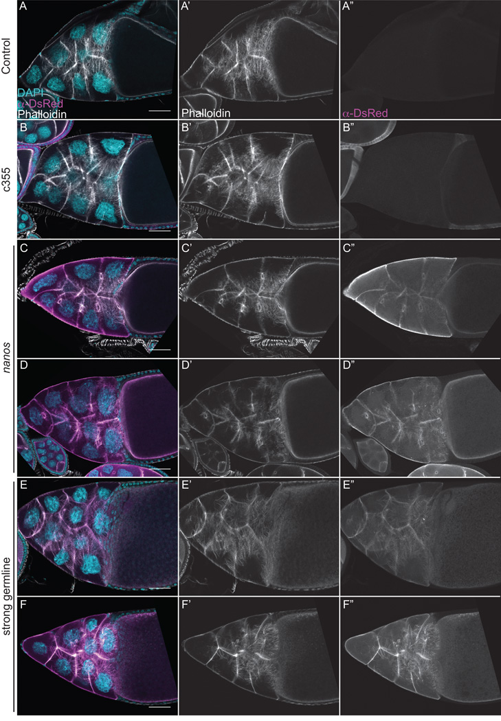
Figure 9. F-tractin-tdTom labels F-actin structures during S10B without causing actin defects.
(A-F”) Maximum projections of 3–5 confocal slices of fixed and stained S10B follicles taken at 20X magnification. (A-F) Merged images: DNA (DAPI) = cyan, F-actin (phalloidin) = white, F-tractin (anti-DsRed) = magenta. (A’-F’) F-actin (phalloidin) = white. (A”-F”) F-tractin (anti-DsRed)= white. (A-A”) F-tractin-tdTom/+. (B-B”) c355 GAL4; F-tractin-tdTom/+. (C-D”) F-tractin-tdTom/+; nanos-VP16 GAL4. (E-F”) Strong germline GAL4 (either mat2MK, mat3, or oskar GAL4) driving F-tractin-tdTom. Somatic expression of F-tractin-tdTom does not alter nurse cell actin remodeling or follicle cell morphology during S10B (B-B” compared to A-A”). Follicles weakly expressing F-tractin-tdTom in the germline exhibit normal nurse cell actin filament bundles and cortical actin deposits (C-D” compared to A-A”). However, the actin filament bundles within the nurse cells are unlabeled (C-C”) or only weakly labeled (D-D”) when F-tractin-tdTom is weakly expressed using nanos-VP16 GAL4. Strong germline expression of F-tractin-tdTom does not cause striking defects in nurse cell actin filament bundles and cortical actin deposits, and actin bundles are labeled, although to varying degrees (E-F”). Scale bars = 50 μm.

