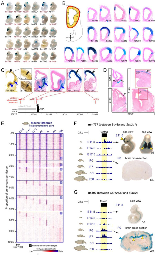
Figure 3. Spatial and temporal specificity of enhancers active in the developing forebrain.
A) Subset of forebrain enhancers with a spectrum of subregional specificities at whole-mount resolution. B) Examples of enhancers with restricted pallial activity in the mouse telencephalon at E11.5. C) Multiple enhancers in the larger region surrounding the Arx gene show subregional forebrain activity patterns that recapitulate endogenous Arx mRNA expression in the mouse forebrain. Notably, enhancer activities show partial spatial redundancy. D) Example of an enhancer with activity across multiple developmental stages, labeling cell populations whose location is consistent with migration from the MGE, through the LGE, to the cortex (white arrows). E) Developmental dynamics of enhancer-associated histone mark H3K27ac at candidate forebrain enhancers analyzed by ChIP-seq across seven stages of brain development. Most sites show temporally restricted H3K27ac marks. F,G) Examples of in vivo validated temporally dynamic enhancer activity in the forebrain, as predicted by temporally dynamic H3K27ac signatures. CP, choroid plexus; Cx, cortex; CxP, cortical plate; DP, dorsal pallium; LGE, lateral ganglionic eminence; LP, lateral pallium; MGE, medial ganglionic eminence; MP, medial pallium; MZ, marginal zone; VP, ventral pallium. A-D modified from Visel et al., 2013. E-G modified from Nord et al., 2013.

