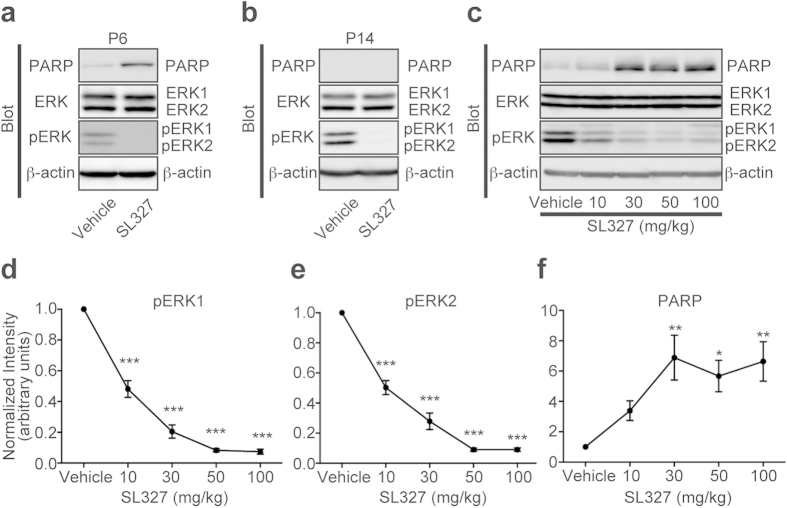
Figure 2. Transient SL327 administration causes apoptosis in the brain with strong age-dependency.
(a) Representative images of western blot analysis using anti-cleaved PARP antibody showing that SL327 administration at P6 caused significant increase of apoptosis in the mouse compared with administration of vehicle (DMSO) (n = 10 for each). (b) Conversely, SL327 did not induce increased apoptosis at P14 (n = 6 for each). (c) Representative images of western blot analysis investigating the effect of each concentration of SL327 (0 (vehicle), 10, 30, 50, and 100 mg/kg). Expression levels of ERK1 and ERK2 were not significantly changed among concentrations tested. (d) The phosphorylation levels of ERK1 were decreased in a dose-dependent manner with saturation above 50 mg/kg of SL327. (e) The phosphorylation levels of ERK2 were decreased in a dose-dependent manner with saturation above 50 mg/kg of SL327. (f) SL327 concentrations above 30 mg/kg caused significant increase of apoptosis in the mice forebrain 6 h after administration. To evaluate expression and phosphorylation, band levels were divided by their corresponding loading internal control (β-actin). All the gels were run under the same experimental conditions. Cropped blots are shown in western blot data. Data are represented as mean ± SEM. *P < 0.05, **P < 0.01, ***P < 0.001 compared with vehicle controls (n = 7 mice for each concentration).

