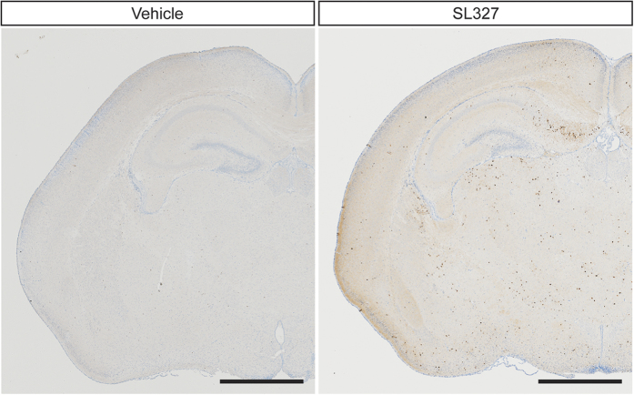
Figure 3. Number of black dots labeled by immunohistochemical staining for activated cleaved caspase-3 was increased in the brain from mice administered SL327 at P6.
Representative light microscope views of coronal sections from the mouse brain 6 h after injection of vehicle or SL327. Black or brown dots represent immunostaining for activated caspase-3. High density of cleaved caspase-3 positive profile was present in mice administered SL327. Mouse brains were removed 6 h after injection. Scale bars: 1 mm.

