
Figure 2.
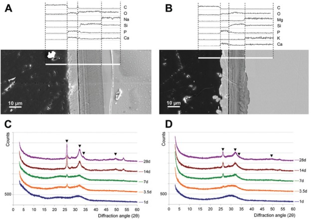
45S5 (A, C) and PC-XG3 (B, D) after storage in simulated body fluid (SBF). A and B show exemplary cross sections of the resulting surface layers after 14 d. Bulk glass is shown on the right side and the embedding material on the left side of the picture. White lines represent the zones of elemental analysis. C and D show x-ray diffraction results after 1 to 28 days of storage. Each diagram was scaled up about 500 counts for easier reading. The marked peaks correspond with carbonate hydroxyapatite (powder diffraction file 00-019-0272, database PDF4+ [2009]; International Centre for Diffraction Data, Newtown Square, PA, USA).