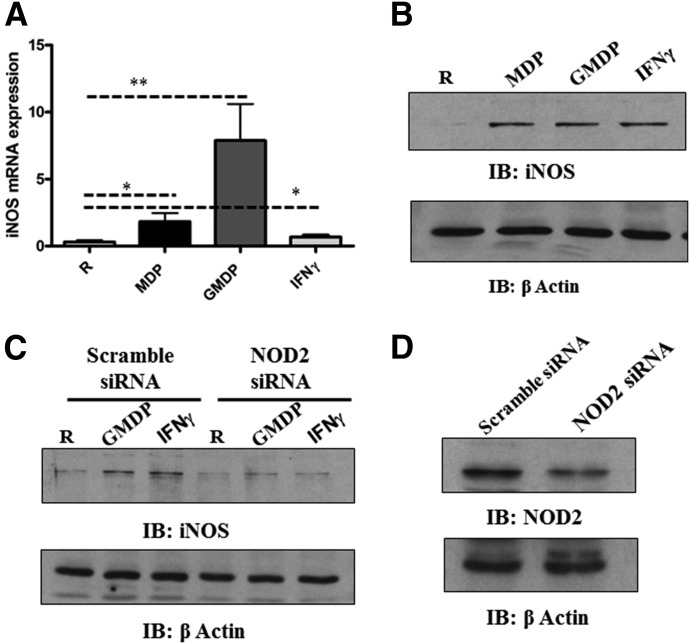
Figure 1. NOD2 activation regulates iNOS expression in human macrophages.
MDM monolayers were treated with MDP (5 μg/ml), GMDP (5 μg/ml), or IFN-γ (10 ng/ml) for 24 h; then, monolayers were lysed with TRIzol for extraction of total RNA or lysed with TN-1 lysis buffer for Western blot. (A) qRT-PCR was used to determine iNOS mRNA levels. Data were normalized to the β-actin gene, and RCN was determined. Shown are cumulative data from 5 independent experiments (mean ± sem; *P < 0.05; **P < 0.005). R, Resting group. (B) Equal amounts of cell lysates were analyzed for iNOS expression or loading control β-actin by Western blot. Shown is a representative experiment from 3 independent experiments. IB, Immunoblot. (C) Scramble or NOD2 siRNA-transfected MDM monolayers were incubated with GMDP (5 μg/ml) or IFN-γ (10 ng/ml) in 1% autologous serum for 24 h. Cells were lysed with TN-1 buffer, and cell lysates were analyzed for iNOS production by Western blot and reprobed for β-actin as a loading control. Shown is a representative experiment of 3 independent experiments. NOD2 knockdown was confirmed by Western blot (D). Shown is a representative experiment of 3 experiments.

