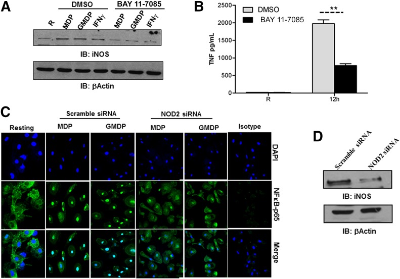
Figure 6. NOD2-mediated NF-κB activation is required for iNOS expression in human macrophages.
Day 5 MDMs were pretreated with the NF-κB inhibitor BAY 11-7085 for 30 min and subsequently stimulated with MDP (5 μg/ml), GMDP (5 μg/ml), or IFN-γ (10 ng/ml) for 24 h. (A) Cells were lysed with TN-1 buffer, and whole-cell lysates were analyzed for iNOS protein levels by Western blot by use of iNOS antibody or β-actin antibody as a loading control. Shown is a representative experiment of 2 independent experiments. (B) In parallel, NF-κB inhibitor-treated cells were stimulated with LPS (50 ng/ml) for 12 h. Cell culture supernatants were used to measure TNF production by ELISA. Shown is a representative experiment (mean ± sd of triplicate samples, n = 2, **P < 0.005). (C) Day 5 MDMs were transfected with scramble or NOD2 siRNA and plated on coverslips after 72 h. Cells were stimulated with MDP, GMDP, or left untreated for 6 h, fixed and permeabilized, and stained with NF-κB-p65 antibody or isotype control, followed by Alexa Flour 488-conjugated secondary antibody. The nuclei were stained with DAPI. Cells were then examined by confocal microscopy for NF-κB p-65 translocation. Shown are representative images from 2 independent experiments. NOD2 knockdown was confirmed by Western blot (D). Shown is a representative experiment of 2 experiments.

