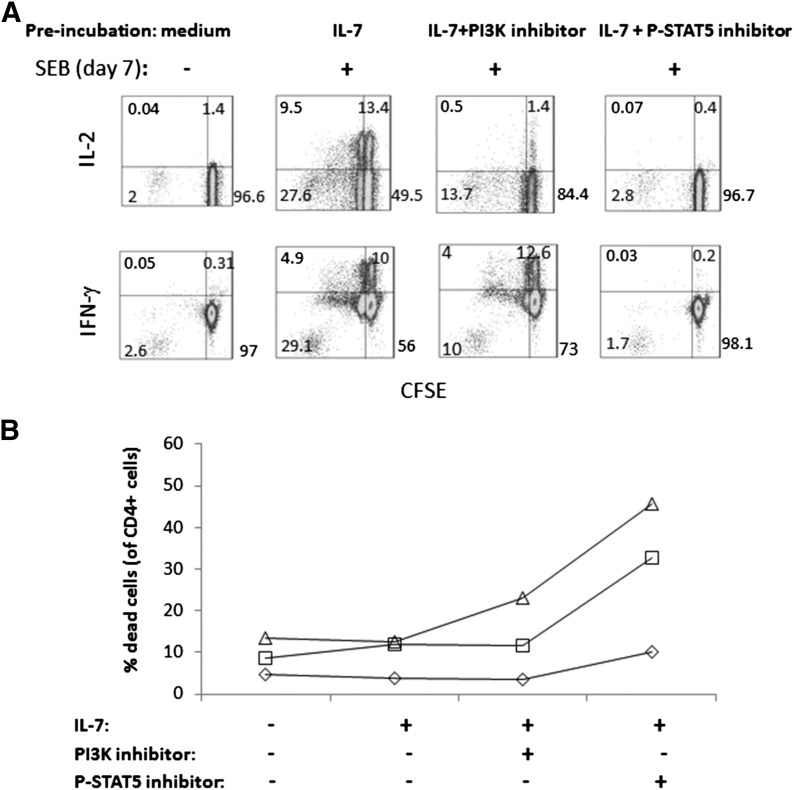
Figure 6. Inhibition of PI3K or STAT5 reduced cell proliferation, decreased functionality, and enhanced cell death in cells treated with IL-7.
(A) CFSE-labeled PBMCs were incubated in medium alone, medium + IL-7, medium + IL-7 + PI3K inhibitor, and IL-7 + STAT5 inhibitor and then assessed 7 days later for proliferation (CFSE dye dilution, x-axis) and response to SEB stimulation (CD40L induction, y-axis). Data are representative of results from 3 different donors. (B) Viability was assessed in the above cultures by gating on forward- and side-scatter to eliminate debris and monocytes, followed by forward-scatter-height versus forward-scatter-area to exclude doublets (not shown). Then, cells were gated on CD4 cells to assess viability dye stain. The percentage of dead CD4+ cells is shown on the y-axis under the various conditions (x-axis). Connected symbols represent data from a single donor. Data from 3 different donors are shown as different symbols.

