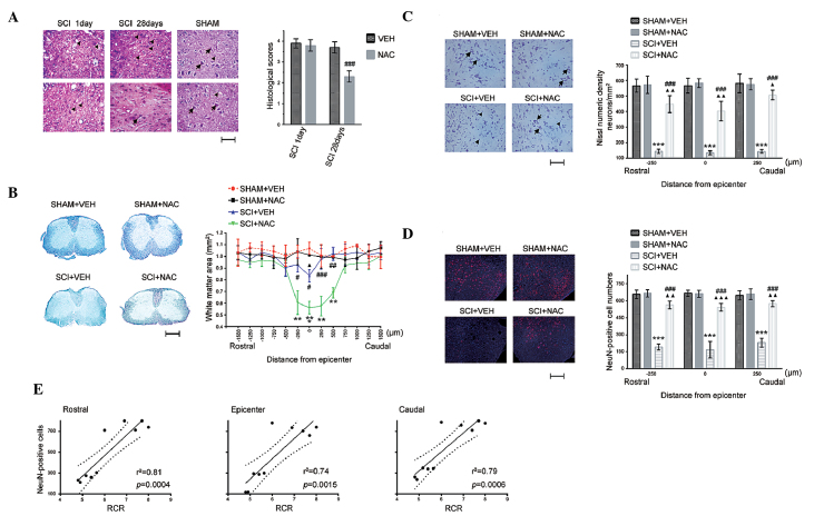
Figure 3.
Effects of NAC on histological changes. Data are presented as the mean ± standard error of the mean of three independent experiments (n=3 per group). **P<0.01, ***P<0.001, SCI+VEH mice versus SHAM+VEH mice; #P<0.05, ##P<0.01, ###P<0.001, SCI+NAC mice versus SCI+VEH mice; ΔP<0.05, ΔΔP<0.01, ΔΔΔP<0.001, SCI+NAC mice versus SHAM+VEH mice. (A) SCI and leukocyte infiltration by hematoxylin and eosin staining at 1 day and 28 days in SHAM and SCI mice treated with NAC or VEH (scale bar=100 μm). Arrows indicate normal neurons; triangles indicate glia. Histological scores were measured for the SCI severity at 1 and 28 days in SCI mice treated with NAC or VEH. (B) White matter sparing by Luxol fast blue staining at 28 days in SHAM and SCI mice treated with NAC or VEH (scale bar=500 μm). Data are expressed as the proportional area. (C) Quantification of Nissl numeric density (neurons/mm2) at 28 days in SHAM and SCI mice treated with NAC or VEH (scale bar=100 μm). Arrows indicate normal neurons; triangles indicate neuron death. (D) Quantification of immunofluorescence staining of NeuN (red) and 4′,6-diamidino-2-phenylindole (blue) at 28 days in SHAM and SCI mice treated with NAC or VEH (scale bar=100 μm). (E) Correlation between mitochondrial RCR and neuronal survival in SCI. NAC, N-acetylcysteine; VEH, vehicle; SCI, spinal cord injury; RCR, respiratory control ratio; NeuN, neuronal nuclei.

